⚠️ 以下所有内容总结都来自于 大语言模型的能力,如有错误,仅供参考,谨慎使用
🔴 请注意:千万不要用于严肃的学术场景,只能用于论文阅读前的初筛!
💗 如果您觉得我们的项目对您有帮助 ChatPaperFree ,还请您给我们一些鼓励!⭐️ HuggingFace免费体验
2025-03-15 更新
X-GAN: A Generative AI-Powered Unsupervised Model for High-Precision Segmentation of Retinal Main Vessels toward Early Detection of Glaucoma
Authors:Cheng Huang, Weizheng Xie, Tsengdar J. Lee, Jui-Kai Wang, Karanjit Kooner, Jia Zhang
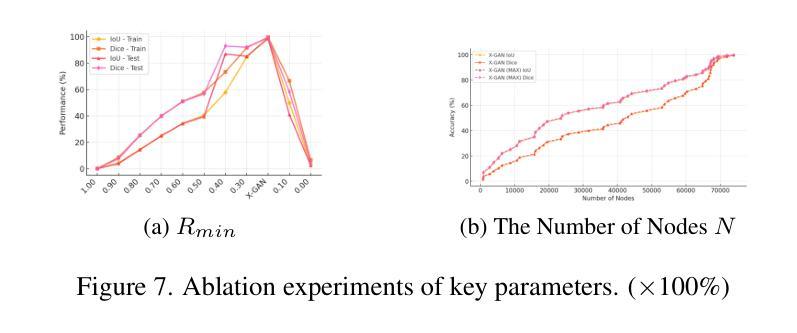
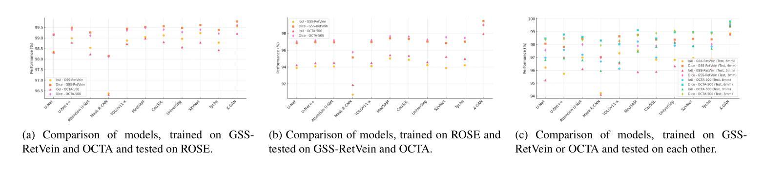
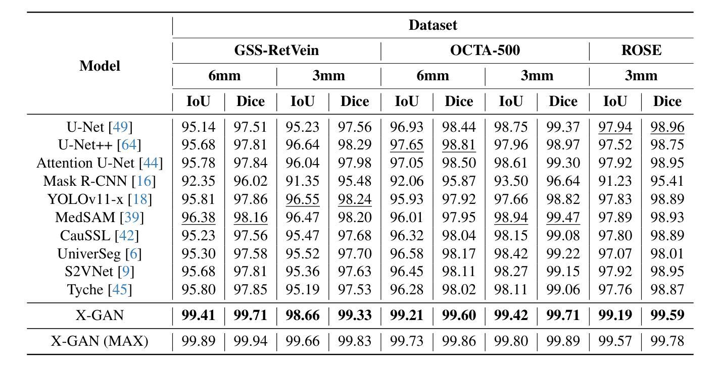
Structural changes in main retinal blood vessels serve as critical biomarkers for the onset and progression of glaucoma. Identifying these vessels is vital for vascular modeling yet highly challenging. This paper proposes X-GAN, a generative AI-powered unsupervised segmentation model designed for extracting main blood vessels from Optical Coherence Tomography Angiography (OCTA) images. The process begins with the Space Colonization Algorithm (SCA) to rapidly generate a skeleton of vessels, featuring their radii. By synergistically integrating generative adversarial networks (GANs) with biostatistical modeling of vessel radii, X-GAN enables a fast reconstruction of both 2D and 3D representations of the vessels. Based on this reconstruction, X-GAN achieves nearly 100% segmentation accuracy without relying on labeled data or high-performance computing resources. Also, to address the Issue, data scarity, we introduce GSS-RetVein, a high-definition mixed 2D and 3D glaucoma retinal dataset. GSS-RetVein provides a rigorous benchmark due to its exceptionally clear capillary structures, introducing controlled noise for testing model robustness. Its 2D images feature sharp capillary boundaries, while its 3D component enhances vascular reconstruction and blood flow prediction, supporting glaucoma progression simulations. Experimental results confirm GSS-RetVein’s superiority in evaluating main vessel segmentation compared to existing datasets. Code and dataset are here: https://github.com/VikiXie/SatMar8.
视网膜主血管结构的变化是青光眼发生和发展的关键生物标志物。识别这些血管对血管建模至关重要,但极具挑战性。本文提出了X-GAN,这是一种基于生成式人工智能的无监督分割模型,旨在从光学相干断层扫描血管造影(OCTA)图像中提取主血管。流程始于空间殖民算法(SCA),该算法可以快速生成血管的骨架并显示其半径。通过协同整合生成对抗网络(GANs)与血管半径的生物统计建模,X-GAN可以快速重建血管的2D和3D表示。基于这种重建,X-GAN在不依赖标记数据或高性能计算资源的情况下实现了近100%的分割精度。此外,为了解决数据稀缺的问题,我们引入了GSS-RetVein,这是一个高分辨率的混合2D和3D青光眼视网膜数据集。GSS-RetVein由于其毛细血管结构异常清晰而提供了严格的基准测试,并且引入了受控噪声以测试模型的稳健性。其2D图像具有清晰的毛细血管边界,而3D组件则增强了血管重建和血流预测,支持青光眼进展模拟。实验结果表明,与现有数据集相比,GSS-RetVein在评估主血管分割方面具有优势。代码和数据集地址为:https://github.com/VikiXie/SatMar8。
论文及项目相关链接
PDF 11 pages, 8 figures
Summary
本文主要介绍了针对青光眼视网膜血管模型构建的X-GAN技术。该技术结合了生成对抗网络(GANs)和血管半径的生物统计建模,能够实现对光学相干断层扫描血管造影图像(OCTA)中的主要血管的快速重建。实验证实,该技术能无标签数据支持且计算资源需求低的情况下达到近乎百分之百的分割准确度。此外,为应对数据稀缺问题,引入了GSS-RetVein数据集,包含清晰的毛细血管结构并加入可控噪声以测试模型稳健性,支持青光眼进展模拟。
Key Takeaways
- 结构变化在视网膜主要血管中作为青光眼发病和进展的关键生物标志物。
- X-GAN是一种基于生成对抗网络(GANs)的无监督分割模型,用于从OCTA图像中提取主要血管。
- X-GAN结合了血管半径的生物统计建模和空间殖民算法(SCA),实现了快速血管重建。
- X-GAN在不依赖标签数据和高性能计算资源的情况下达到了接近百分之百的分割精度。
- 为应对数据稀缺问题,引入了GSS-RetVein数据集,为评估血管分割提供了严谨的基准测试。数据集包括清晰毛细血管结构的二维和三维图像,支持青光眼进展模拟。
点此查看论文截图